Cut your study time.
Test yourself.
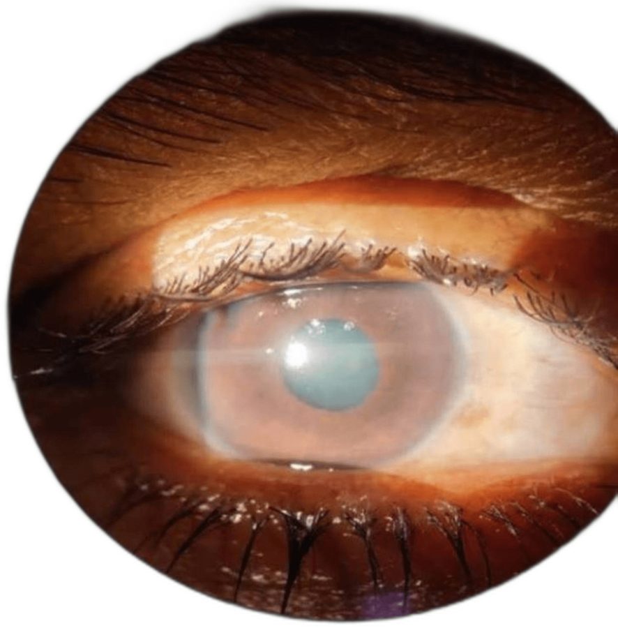
Thool AR, Selukar KV, Daigavane SV (2023), Cureus, doi:10.7759/cureus.34621 · CC BY 4.0 · Converted.
An adult comes in with a painful right eye and haloes; the slit lamp shows the picture above. The attack is broken with drops and a laser iridotomy. Two days later the cornea is clear and the pressure is 9 mmHg without drops. What is the next step?
إجابة غير صحيحة. الصحيحة: B.
A low pressure after an attack is not an open angle; look at the angle again.
After an attack the ciliary body is ischaemic and makes less aqueous, so the pressure can stay low for weeks whatever the angle is doing. Pressure is therefore a poor guide to the angle; repeat or serial gonioscopy shows how much of it has reopened and how much is held closed by synechiae. The fellow eye usually shares the anatomy and is treated with an iridotomy, not watched.
Review before you forget.
البطاقات
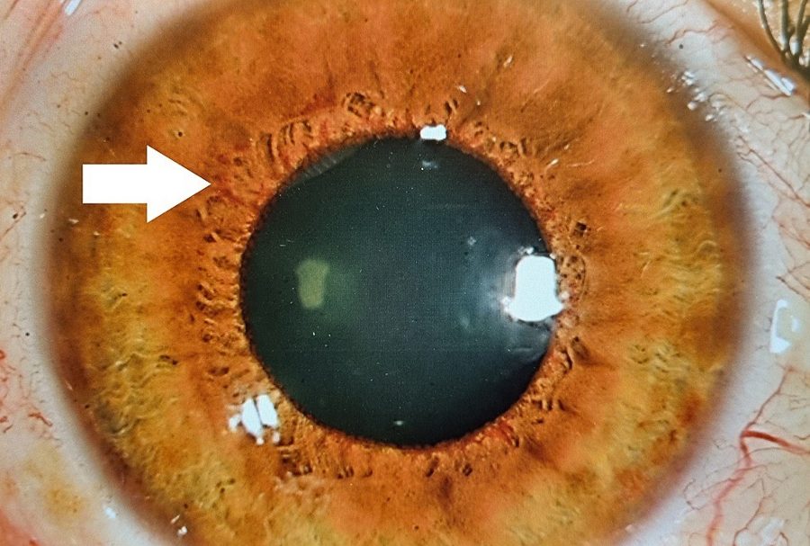
What is at the arrow, and where must you look next?
اعرض الإجابة
New vessels on the iris (rubeosis), starting at the pupil margin.
VEGF from ischaemic retina drives vessels on the iris and in the angle; a fibrovascular membrane then pulls the angle shut — neovascular glaucoma. Look at the angle by gonioscopy and at the retina for the cause: diabetic retinopathy, a vein occlusion or ocular ischaemia. Treat the ischaemia: PRP, with anti-VEGF to act faster.
المصدر · ص 172–174Rubeosis iridis is seen at the pupillary margin area, almost 360 degrees. Zakaria et al. (2023), Cureus, doi:10.7759/cureus.43303 · CC BY 4.0 · Converted.
Take notes with your pen.
A low pressure after an attack is not an open angle — ciliary shutdown can keep it low for weeks; gonioscope again.
Check the angle on day 2.
Know the sign by its picture.
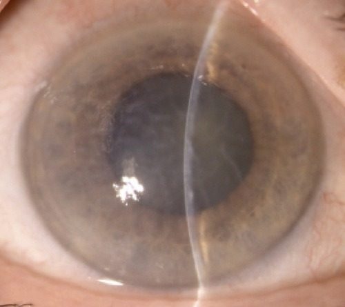
Vivarelli C, Pellegrini M, Parmeggiani F, et al. (2026), Case Rep Ophthalmol Med, doi:10.1155/crop/8847063 · CC BY 4.0 · Converted.
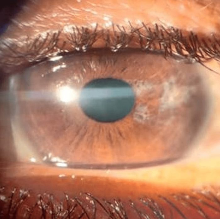
Thool AR, Selukar KV, Daigavane SV (2023), Cureus, doi:10.7759/cureus.34621 · CC BY 4.0 · Converted.
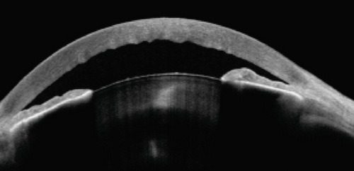
Vivarelli C, Pellegrini M, Parmeggiani F, et al. (2026), Case Rep Ophthalmol Med, doi:10.1155/crop/8847063 · CC BY 4.0 · Converted.
Search by symptom or diagnosis.